Where do spinal fractures most commonly occur?
The spine is the central support structure of the body that transfers body weight to the lower extremities and maintains balance and posture. It serves a critical protective function as it encloses the spinal cord, which carries signals between the brain, organs, and limbs.
The spine consists of seven cervical vertebrae (neck), twelve thoracic vertebrae (upper back), five lumbar vertebrae (lower back), five sacral vertebrae (pelvis), and three to four coccygeal vertebrae (tailbone). Vertebrae are numbered from the top of the spine downward, so the seventh cervical vertebra is referred to as C7.
The most commonly injured regions are the cervical spine and the thoracolumbar junction (the area connecting the thoracic and lumbar spine). The cervical spine is vulnerable because it connects the rigid torso to the relatively heavy head with considerable flexibility. The thoracolumbar region is prone to injury because it represents the longest segment of the spine and undergoes the most movement.
Spinal fractures typically result from strong external forces such as falls or motor vehicle accidents, and their frequency has increased with industrialization. Additionally, as life expectancy increases, osteoporotic compression fractures caused by relatively minor trauma in elderly patients are becoming increasingly common.
What immediate steps should be taken when encountering a patient with spinal injury?
Emergency care for spinal injury begins at the accident scene. First, vital signs such as breathing and circulation should be assessed to determine if the patient's life is in immediate danger, and appropriate emergency measures should be taken. When transporting a patient with suspected spinal injury, a cervical collar should be applied, the patient should be placed on a firm board, and the torso should be supported on both sides with sandbags to prevent movement. This reduces the risk of secondary neurological injury from fracture site movement in cases of unstable fractures. Once the patient arrives at the hospital, evaluation for other significant injuries should be performed, and the patient should be managed assuming spinal injury exists until spinal injury is definitively ruled out.
What symptoms appear with spinal fracture?
Pain at the fracture site is the most basic and common symptom. When there is damage to spinal nerves—specifically the spinal cord or nerve roots—limb pain or paralysis may occur. Following relatively severe trauma such as falls or motor vehicle accidents, spinal injury must be investigated regardless of whether the patient reports pain.
In cervical injuries where there is only fracture or dislocation without damage to the spinal cord, patients may experience neck or shoulder pain and instability with motion. When instability is present, neck movement may significantly worsen pain. Patients who initially believe the injury is minor may think the pain will resolve on its own, but persistent pain should raise suspicion of instability-related discomfort.
Although it is fortunate if there is no neurological damage, when a fractured vertebra injures the spinal cord inside it, sensation in the arms or legs may become dull or absent, paralysis may develop causing muscle weakness or complete loss of movement, and bladder dysfunction may prevent normal urination. If there is a fracture between the first and fourth cervical vertebrae with associated spinal cord damage, paralysis of the respiratory muscles may develop, resulting in respiratory failure. It is essential to assess whether neurological (spinal cord and nerve root) damage has occurred and to what extent in patients with spinal fractures, as this is critically important for surgical decision-making and prognosis.
What other areas of the body may be injured along with the spine?
When trauma is severe enough to cause spinal damage, there is a high likelihood of concurrent head trauma, internal organ injury, or other skeletal injuries. Assessment for related symptoms is important. Assessment of life-threatening conditions such as respiration and circulation takes priority, and careful examination of the head, chest, abdomen, and extremities is necessary in addition to the spine. Other limb fractures commonly associated with spinal fractures include calcaneal (heel bone), tibial (shin bone), and wrist fractures.
When traumatic disc herniation occurs, symptoms are similar to typical disc herniation. Middle-aged and older patients may have degenerative changes in the cervical vertebrae and discs even without prior symptoms, so even minor trauma can cause severe symptoms.
What tests are performed?
Patients with suspected spinal or spinal cord injury undergo X-ray imaging immediately. If suspicious lesions are identified or detailed imaging is needed, CT (computed tomography) or MRI (magnetic resonance imaging) scans are performed. CT provides clear diagnosis of fractures and is superior to MRI in detecting bone fragments protruding into the spinal canal. MRI imaging most accurately shows whether blood has entered the spinal cord, the degree of swelling, and whether the disc is ruptured. MRI can also detect ligament injuries that other tests cannot identify. Combined CT and MRI imaging of the injured spine provides the most accurate diagnosis.
What treatment options are available?
1) Cervical (neck) injuries
In patients with cervical fracture where the fractured bone is not significantly displaced, spinal instability is absent, and there is no neurological damage, wearing a cervical collar and reducing pain with anti-inflammatory analgesics while maintaining rest typically results in good recovery. If there is spinal instability and fractured bone fragments are compressing the nerve, surgery is needed to relieve the nerve compression and insert fixation devices to stabilize the spine. Even with internal metal fixation, a cervical collar or external fixation device may be needed after surgery.
For fractures at cervical levels 1 and 2 where spinal instability is present but the spinal cord is uninjured, some patients may wear a rigid external fixation device (brace) and maintain rest for up to 6 months. Because long-term external fixation has limitations and surgery may be needed if the fracture fails to heal properly, early surgical fixation is increasingly preferred.
For fractures in the lower cervical spine, if spinal instability or misalignment is present, traction devices that pull the head to restore normal alignment are applied, followed by surgery to rigidly fix the spine. When traumatic disc herniation compresses the nerve or fractured bone fragments or misaligned vertebrae compress the nerve, surgery is needed to decompress the nerve and stabilize the spine.
2) Thoracolumbar (mid-back and lower back) injuries
The goals of treating thoracolumbar spinal fractures are to maintain life, protect neurological function, restore proper spinal alignment to achieve stability, enable early mobilization and rehabilitation, and facilitate return to society.
Fractures without neurological symptoms and showing stability are treated conservatively (non-surgically), while those with instability or risk of neurological injury are treated surgically.
The goal of surgical treatment is to firmly fix fractures and deformities using metal screws and similar devices to promote neurological recovery and enable early mobilization.
Surgery is indicated in three situations: first, when lower extremity paralysis from neurological injury is present and imaging shows fractured bone compressing the nerve; second, when the fracture site is unstable, risking deformity or additional paralysis if left untreated, requiring fracture fixation; third, when the vertebral body is severely collapsed from the fracture.
The timing of surgery is decided by weighing how severely the nerves have been injured together with how unstable the fracture site is; there is no fixed waiting period of a set number of days as was once taught. Rather, the trend is toward stabilizing the fracture earlier once surgery is judged to be necessary. Open fractures (compound fractures) or progressive neurological paralysis are better treated with emergency surgery.
Surgical methods include fixing the spine from the front or back using metal plates or rods, and when necessary, directly removing bone fragments compressing the nerve or grafting bone to promote fracture healing.
Non-surgical methods include prolonged bed rest, posterior extension plaster fixation of the thoracolumbar region, bed rest followed by brace wear, and immediate mobilization without bracing from the start. The choice depends on fracture type, location, patient age, and presence of associated injuries.
Current treatment trends favor minimizing bed rest duration. Once fracture site pain is adequately relieved and the patient can sit with minimal discomfort, a brace is applied and mobilization is begun. Braces are typically worn for at least 3 months, after which X-rays are obtained without the brace. If no further collapse or deformity is seen at the fracture site, the brace is discontinued. Subsequently, spinal muscle strengthening exercises are beneficial to recover from muscle weakness due to immobility.
If despite adequate brace wear progressive deformity or abnormal motion develops at the fracture site with associated pain, surgical treatment becomes necessary.
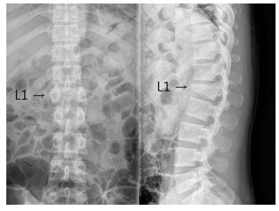
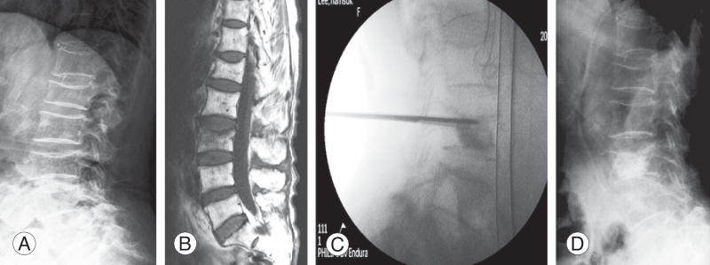
1. Example of a burst fracture of the first lumbar vertebra (L1) in a 35-year-old male. The fractured vertebral body is compressed compared to the normal vertebral bodies above and below, and a kyphotic deformity with anterior angulation of the fracture is also present.
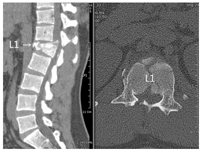
2. In burst fractures, bone fragments from the fractured vertebral body are driven into the spinal canal, compressing the spinal nerve. Computed tomography more clearly shows the intrusion of bone fragments into the spinal canal.
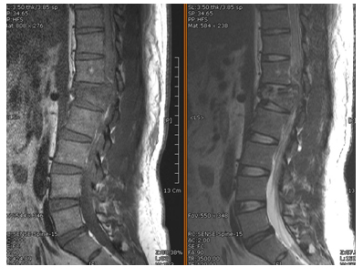
3. MRI imaging clearly shows compression and damage to the nerve root from this displacement.
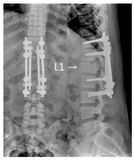
4. Postoperative image following surgical treatment. Pedicle screws are inserted into the segments above and below the fractured vertebra, and metal rods are connected like bridges to correct the fractured vertebral body.
How does osteoporotic spinal compression fracture differ from general fractures?
Unlike healthy individuals, osteoporotic compression fracture refers to compression deformity of weakened vertebrae from relatively minor trauma. Sometimes fracture occurs from such minor trauma that the patient may not even remember the injury.
Symptoms differ from acute lower back pain; localized spinal pain persisting 1-2 weeks after trauma should raise suspicion. Unlike typical leg pain, thoracic or abdominal pain may accompany the spinal pain.
What tests are performed when osteoporotic spinal compression fracture is suspected?
With a history of trauma and pain, radiological imaging should be obtained. Diagnosis proceeds with X-ray imaging as with general fractures, with CT (computed tomography), MRI (magnetic resonance imaging), or bone scan performed as needed. CT is good for assessing vertebral status but can make distinguishing acute from chronic fractures difficult. MRI is excellent for assessing the spinal cord, disc, and bone status and can determine the timing of injury, though it is costly. For elderly patients, MRI is very useful for differentiating other conditions such as metastatic cancer to the spine. Bone scans can assess multiple systemic fractures but have limited resolution.
How are osteoporotic compression fractures treated?
Once osteoporotic compression fracture is diagnosed, rest on a relatively firm surface with a soft mat is recommended for a short period. Conservative treatment is initiated first, with medication and physical therapy typically resulting in fracture healing within 2-3 months. In this case, pain resolves but vertebral deformity persists.
However, if pain has not improved 2 weeks after fracture, the patient is 80 years or older, osteoporosis is severe, or complications from prolonged bed rest are anticipated (such as diabetes or heart disease), vertebroplasty may be performed to allow early mobilization. Vertebroplasty and balloon kyphoplasty do not require general anesthesia and are quick and safe, making them among the most widely used methods. Pain typically improves immediately after the procedure, and balloon kyphoplasty can potentially restore some of the compressed vertebral height. However, if the spinal fracture is severe with bone fragments compressing the nerve or nonunion with persistent instability, vertebral body fixation surgery may be needed. In such cases, detailed consultation with an orthopedic spine specialist is necessary.
Medical review: Spine Osteoporosis Research Society · Provided by the Korean Society of Spine Surgery