What kind of surgery is endoscopic spinal surgery?
When patients visit the hospital with lower back and leg pain and receive a disc disease diagnosis, various treatments are recommended. Perhaps starting with medication, or nerve block treatment is recommended. Of course, in most cases these conservative non-surgical treatments improve symptoms. However, if severe pain persists despite such non-surgical treatment, detailed imaging tests (CT or MRI) are recommended, and after testing you might hear the shocking words: 'Your disc has ruptured. You need surgery.' At such times, one of the natural questions in patients' or guardians' minds is: 'How is the surgery done? Do you open it up or use laser?'
'Do you open it up?' refers to the traditional method of 'open surgery' where skin is incised and operated, while 'Do you use laser?' refers to 'minimally invasive endoscopic techniques' for surgery. This section addresses the curiosity about that 'Do you use laser?' question.
First, let's understand what 'laser' means as patients and general public ask doctors. Medical equipment called 'laser' includes Holmium-YAG Laser, CO2 Laser, and others. Particularly, H-YAG Laser side-firing systems used in spinal surgery are so expensive and have limited application that they are not commonly used equipment. Laser used in spinal endoscopic surgery mainly refers to High Radiofrequency Electrocoagulating System, thus there is a slight difference between 'laser' discussed at surrounding hospitals and the 'laser' patients imagine. Eventually, 'lumbar disc laser surgery' is more accurately called 'spinal endoscopic micro-surgery for lumbar disc utilizing high-frequency radiofrequency electrocoagulation as surgical aid'.
How is endoscopic surgery performed?
Second, let's answer 'How is endoscopic surgery performed?' Recent highly developed optical technology and endoscope manufacturing technology have led to development of various spinal endoscopic equipment and surgical tools. Ultimately, endoscope development has divided disc surgery into three categories: 1. Cases where both open and endoscopic surgery are possible, 2. Dramatic cases where complicated or difficult cases in open surgery are solved more simply endoscopically, and finally 3. Rare cases that remain unsolved requiring complex surgery. Surgical methods broadly include the interlaminar approach from the posterior (back) like conventional surgery and the transforaminal approach from the lateral (side).
Interlaminar approach (posterior)
This method approaches using endoscopy as in conventional open surgery and directly removes the disc under endoscopic visualization. Therefore, indications are the same as conventional surgery.
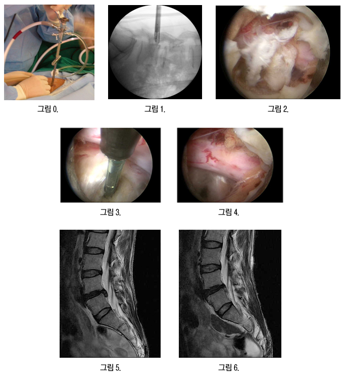
Figure 0 shows the surgical scene, Figure 1 is an image taken during surgery with the C-arm image intensifier, and Figure 2 shows the herniated disc around the exposed nerve root. Figure 3 shows the disc being cleared with radiofrequency, Figure 4 shows the nerve root now freed, and Figures 5 and 6 are the pre- and post-operative MRI, confirming that all of the herniated disc has been removed.
Transforaminal approach (from the side)
This method is performed when the herniated disc occurs not in the typical location but in the outer side of the spinal canal or in the intervertebral foramen. In cases where conventional open methods would be difficult and complex or might require spinal screw fixation due to joint damage, discs can be removed very simply using endoscopy.
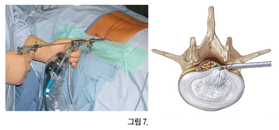
Figure 7 shows the surgical scene and is also a diagram of the surgical approach.
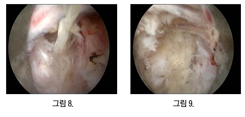
Figure 8 shows the nucleus pulposus exposed outside the torn annulus fibrosus, and Figure 9 shows that extruded nucleus (red line) brought into view through the endoscope.
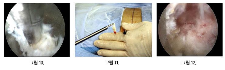
Figures 10 and 11 show the herniated nucleus pulposus being removed through the endoscope, and Figure 12 is the endoscopic view after the nucleus has been cleanly removed.
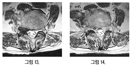
Figure 13 is the pre-operative MRI and Figure 14 the post-operative MRI, confirming that the herniated nucleus (red circle) has been completely removed.
What is endoscopic surgery through two portals (biportal endoscopy)?
The methods described so far use a single channel through which the endoscope and the surgical instruments pass together (single-portal, or uniportal, endoscopy). In addition to these, biportal endoscopic spine surgery (UBE), which makes two small openings, has been widely used since around 2010. It is a method that spine surgeons in Korea have researched and developed extensively. The endoscope is inserted through one opening to look inside, while surgical instruments are inserted through the other opening to carry out the treatment. Because the viewing channel and the working channel are separate, the instruments can be moved far more freely, and the instruments already used in conventional spine surgery can be used as they are.
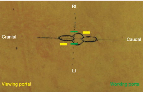
For this reason it is used not only for lumbar disc herniation but also for spinal stenosis, where bone and ligament are trimmed away to widen the passage the nerves run through, and results comparable to surgery performed under the surgical microscope have been reported. It requires one more opening than the single-portal method, but compared with conventional surgery that opens the skin widely, there is less tissue damage and less post-operative pain, and the return to daily life is faster. Rather than one method being better than the other, the appropriate method is chosen according to the location and size of the herniated disc and the degree of stenosis present alongside it.
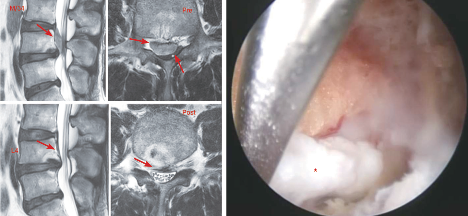
We have now reviewed two representative endoscopic surgical methods for lumbar disc herniation. Endoscopic disc surgery cannot solve all disc diseases, but in some cases can provide surgical results comparable to conventional open surgery, and in rare cases can achieve surgical results not previously possible with conventional open surgery. However, endoscopic disc surgery is not a spectacular laser show; the surgeon's extensive experience and effort are absolutely necessary in this field. Important is that it must be performed with appropriate indications, so careful approach will be necessary.
Medical review: Minimal Invasive Surgery Research Society · Provided by the Korean Society of Spine Surgery