What is internal disc disruption? How is it different from disc herniation?
Internal disc disruption is a syndrome in which persistent, severe lower back pain is caused by abnormalities in the internal structure or metabolic function of the disc with minimal morphological changes in the disc.
Internal disc disruption means there is a problem inside the disc. There is debate among scholars about distinguishing this from disc degeneration that simply occurs with aging. Unlike disc herniation where the disc bulges into the spinal nerve space, directly irritating the nerve and causing pain, in internal disc disruption the disc remains in place but pain still develops in the lower back. The exact mechanisms by which this condition causes pain are speculated to be various. These include: chemical substances released from the ruptured nucleus pulposus inside the disc that irritate microscopic nerves distributed in the membrane surrounding the disc; irritation of the posterior branch of the spinal nerve (called the sinuvertebral nerve), which causes lower back pain; or immune reactions in the body that trigger lower back pain.
Additionally, internal disc disruption often involves loss of water and protein content from inside the disc, resulting in loss of elasticity. Unable to withstand the constantly applied loads, the disc membrane (annulus fibrosus) often develops tears. Like a tire that absorbs impact well when it has adequate air pressure but tears and cannot absorb impact when air-depleted, problems with this impact absorption mechanism lead to pain when the disc cannot properly absorb loads.
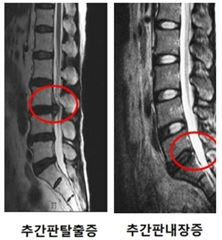
How is internal disc disruption distinguished from disc herniation?
Internal disc disruption has low back pain as the main symptom. It is difficult to bend or sit, and on X-ray or MRI the disc appears black but the shape usually looks normal. In contrast, disc herniation (the condition commonly called a slipped disc) shows the disc shape bulging backward on CT or MRI imaging. Symptoms include primarily buttock pain or pain radiating down the back or front of the leg from the buttock, and pain changes depending on movement and leg position.
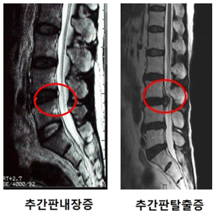
What are the symptoms of internal disc disruption?
Symptoms include persistent lower back pain that worsens when sitting or bending forward, and may be accompanied by buttock or lower extremity pain. When pain is localized deep in the lower back, it does not improve well even with rest. Over time, the condition worsens and is aggravated by work or exercise.
A characteristic feature is inability to sit for prolonged periods and difficulty straightening the back when rising. Symptoms worsen when bending forward. Additionally, because patients do not understand the cause and the pain persists, depression or stress-related weight loss can occur.
What causes internal disc disruption?
The main cause of pain is known to develop after trauma, but it can also develop without trauma. It can occur following external injury from strong forces, such as motor vehicle accidents, or from prolonged strain from sustained labor. Or, such as in workplace accidents where someone falls from height, or after a simple back strain, or after the back twists, or after a fall on the buttocks, if chronic lower back pain develops that does not improve, particularly if that pain is worse when sitting or bending forward, internal disc disruption should be considered.
When the inside of the disc is damaged by trauma, irritative chemical substances are generated to heal the injury. These substances irritate surrounding nerves or trigger immune responses causing pain. Additionally, when the outer membrane of the disc, which is richly innervated with nerve tissue, ruptures and abnormal new nerve growth occurs, there is increased sensitivity to pain compared to normal circumstances.
How is internal disc disruption diagnosed? Can it be diagnosed by MRI?
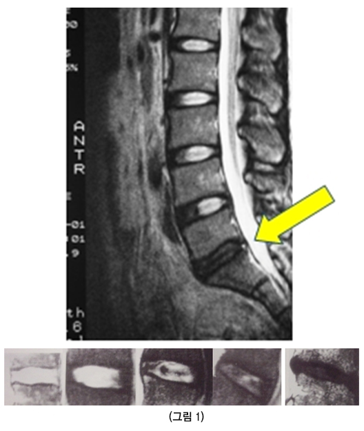
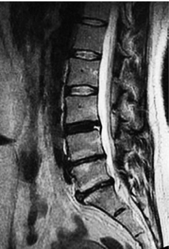
Diagnosis begins with taking a thorough medical history from the patient. If the patient has a history of past injury (though this may be absent) followed by chronic low back pain that does not improve, with symptoms worsening when sitting or bending forward, this condition should be suspected and MRI is recommended. As seen in the image, as water and protein content is lost from inside the disc, elasticity is lost. Depending on the degree of loss, the disc becomes darker on specific MRI sequences (Figure 1).
On MRI, if the disc appears dark and additionally shows signs of rupture in the membrane surrounding the disc appearing white on MRI (arrow), internal disc disruption should be suspected. However, diagnosis based on this alone is difficult. In some cases, internal disc disruption is diagnosed even when the disc is not dark but appears white. Therefore, the next diagnostic step involves performing a discography.
What is discography and how is the diagnosis made?
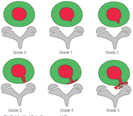
In this procedure, a needle is inserted into the disc and medication is injected. The degree of rupture inside the disc appears as an image. As shown in the figure, it can range from cases where the nucleus pulposus appears neatly like cotton (Figure 2) to cases where the nucleus pulposus rupture extends all the way to the outer annulus fibrosus. However, compared to the imaging appearance of the degree of rupture, the pattern of pain reproduction in the patient is more important for diagnosis. Typically divided into 4 types of response, if the patient's everyday lower back pain is similarly or identically reproduced, the diagnosis of this condition can be established. Even if there is pain, if it differs from the patient's usual low back pain or if no pain is felt at all, regardless of how much the imaging shows disc rupture, the imaging findings have no diagnostic significance.
Discography is, however, a test to decide on carefully. Reports indicate that in roughly 25% of cases a disc that is not actually the painful one still tests as painful, so its accuracy is debated, and some professional societies recommend against diagnosing a disc as the source of pain on this test alone. There are also long-term follow-up studies suggesting that inserting a needle into a disc may itself accelerate disc degeneration. For these reasons it is performed only when other causes have been thoroughly examined and the test is judged truly necessary.
What happens if lower back pain is left untreated? Regarding treatment of internal disc disruption?
Acute lower back pain typically recovers within 2 weeks. About 7% can become chronic, and about one-third of those who develop chronic pain experience sufficient pain that everyday activities or work is difficult.
There are few reports on outcomes when internal disc disruption is left untreated. However, some reports indicate that about 68% of patients improve, but the remainder experience disability in daily life due to pain. In patients middle-aged and older, spontaneous symptom improvement can occur over time. However, the natural history in younger people is not well known.
Treatment begins with general methods. Pain and anti-inflammatory medications can be taken to block chemical substances released from the ruptured disc, epidural injections can be administered, and physical therapy and exercise therapy can be combined. If symptoms do not improve with the above methods, simple procedures such as nucleoplasty or radiofrequency ablation (Figure 3) can be attempted. If there is no improvement despite prolonged treatment, surgery may be considered.
Before deciding on a procedure or surgery, it is important to give exercise therapy and a rehabilitation program a proper trial. Many studies report that intensive exercise and rehabilitation to strengthen the back and abdominal muscles alone leaves many patients doing about as well as those who had surgery.
▶ SpineTube | Strength training when you have a disc problem? ↗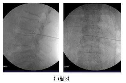
When is surgery absolutely necessary?
If lower back pain has persisted for more than 1 year, has worsened at least once a month, has prevented work for more than 4 months, shows no improvement despite at least 6 weeks of non-surgical methods, and discography reproduces the pain, surgery can be considered. However, diagnosis of this condition should be done carefully, and there is room for debate. Sufficient differentiation from typical lower back pain is necessary, and conservative non-surgical treatment and observation over a sufficient period is advisable.
What surgical methods are available for internal disc disruption?
There are many surgical methods available. The key to surgical treatment is carefully and accurately identifying the disc causing pain and then repairing or removing it. Methods are broadly divided into minimally invasive techniques (without skin incision) and surgical techniques requiring skin incision.
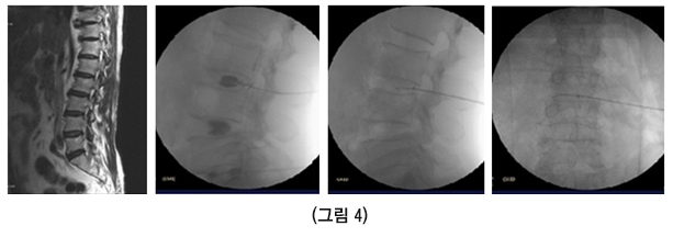
Minimally invasive methods include techniques that use heat to deform and strengthen the nucleus pulposus and annulus fibrosus that cause pain. Nucleoplasty or radiofrequency ablation (Figure 4) can be attempted, but they have not been found to have high success rates and limited usefulness. Therefore, it is very important to make an accurate diagnosis and carefully exclude other causes before performing these procedures, and they should be performed cautiously.
Figure 4 shows an example of a male patient in his late 60s who suffered from lower back pain for several years and showed improvement after undergoing radiofrequency ablation based on MRI and discography findings.
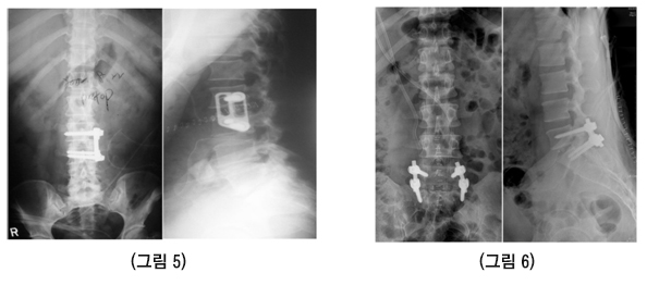
Another surgical method involves removing the problematic disc and then performing fusion surgery by implanting bone into a cage (an artificial spacer device) in the space previously occupied by the disc, fusing the adjacent vertebrae without motion. Success rates for this surgical method are reported to vary from 65-95% among scholars. Accurate diagnosis remains important. This fusion can be performed as anterior fusion surgery, where the abdomen is opened and access is achieved through the front of the disc (Figure 5), or posterior fusion surgery, where the back is incised and access is from behind (Figure 6).
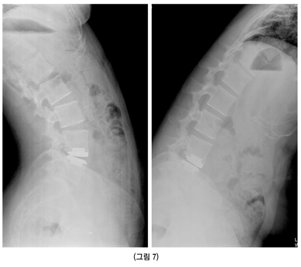
Recently, as interspinous fusion with bone implantation has been found to concentrate stress on the vertebrae above and below the fused level over time, potentially causing additional problems in adjacent areas, an artificial disc has been introduced that preserves the original disc motion instead of fusion. However, this is still considered experimental (Figure 7).
Medical review: Lumbar Spine Research Society · Provided by the Korean Society of Spine Surgery