When a cervical disc herniation or ossification of the posterior longitudinal ligament compresses the nerves of the neck (the spinal cord or the nerve roots) and causes myelopathy or radiculopathy, surgery can be considered if conservative treatment fails to improve the symptoms or they grow worse. Deciding the right timing and the right method is not always easy. Good results follow when an experienced orthopedic spine specialist performs the operation best suited to the problem.
What are the types of surgical procedures available?
An operation that simply removes the structure pressing on the nerve is called a simple decompression. When, after decompressing, bone is fused between the spinal segments to hold them still and give stability, it is called decompression and fusion. These operations are performed from the front or from the back, depending on where the nerve is compressed, on the symptoms, and on which approach is more convenient and safer.
What is simple decompression?
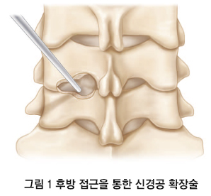
As described above, simple decompression is an operation that simply removes the structure pressing on a nerve root or the spinal cord and causing symptoms. Because it does not fix the spinal segment, it preserves more neck motion after surgery than a fusion does. When a nerve root is compressed, the structure pressing on it (a herniated disc or a bone spur) can be removed from the front or from the back, decompressing the root and relieving the symptoms (Figure 1).
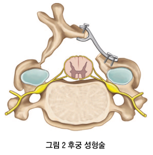
When the spinal cord is compressed, the lesion pressing on it (a bone spur, ossification of the posterior longitudinal ligament, and so on) can be approached from behind with a laminectomy or a laminoplasty. A laminectomy removes the lamina at the back of the vertebra, decompressing the cord so that it is no longer pressed on. After a laminectomy, complications can arise: the cervical spine may bend forward into a kyphotic deformity, or scar tissue may thicken around the dura and press on the cord again, particularly when several levels have been operated on. Laminoplasty is sometimes chosen to prevent these problems (Figure 2). To hold the lamina open after it has been lifted and expanded in a laminoplasty, sutures, wire, autograft, allograft, or small metal plates may be used.
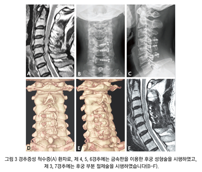
Laminoplasty was once performed as a matter of course on all five levels from C3 to C7, but in recent years C3 and C7 are sometimes only partly released rather than fully opened, to keep postoperative neck pain to a minimum, and at C3 a full laminectomy is sometimes performed instead of a laminoplasty, to prevent loss of motion after surgery (Figure 3).
Neck pain can appear or worsen after posterior cervical surgery, so this deserves attention in patients who already have neck pain before the operation.
What is decompression and fusion?
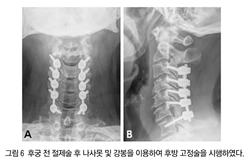
Decompression and fusion means removing the lesion that is pressing on the nerve and then fusing bone between the spinal segments to hold them still and give stability. For a disc herniation or ossification of the posterior longitudinal ligament involving one or two segments, the most commonly performed operation is anterior cervical discectomy and fusion (Figure 4). The cervical spine is approached from the front, the disc is removed to decompress the nerve, and a bone graft is placed between the vertebrae to fuse them. There is some disagreement, but when weakness before surgery is marked or MRI shows severe compression of the nerve root, an uncinate process resection is performed at the same time, burring or cutting away the bone spur and the uncinate process that are pressing on the root. Because this step carries a considerable risk of nerve injury and bleeding and calls for practiced technique, such bone removal is best done by an orthopedic spine specialist. Autologous iliac bone from the patient was used for fusion in the past; a variety of materials is now available, and combining the fusion with metal plate fixation, so that the patient can move sooner and the bone unites faster, is the trend in developed countries.
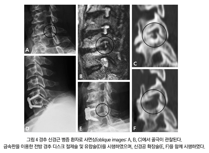
When the spinal cord is compressed from the front and causing myelopathy, and removing the disc alone will not decompress it adequately, an anterior corpectomy and fusion may be performed (Figure 5).
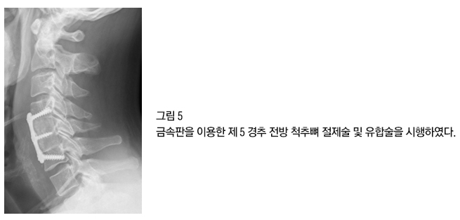
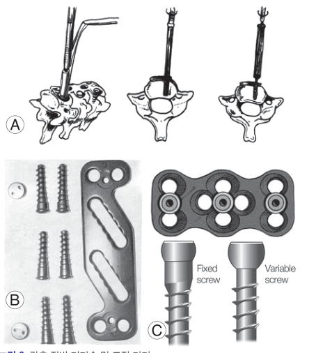
If there is a risk of kyphosis and instability developing after a posterior laminectomy, or if either is already present before surgery, fusion and posterior instrumentation may be added (Figure 6).
What is artificial disc replacement surgery?
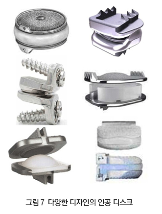
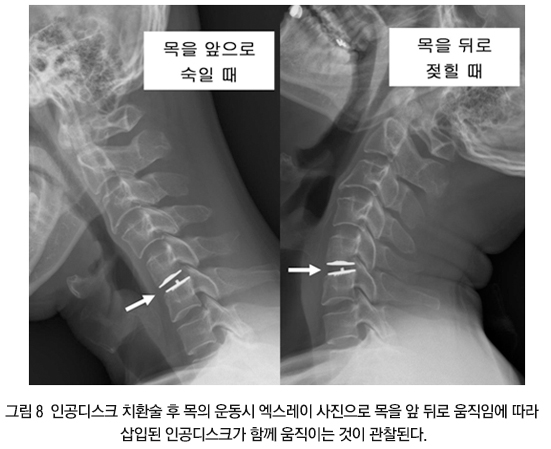
An artificial disc is a device made to imitate the human cervical disc and to be inserted between the vertebrae so that the segment can still move after surgery (Figure 7). After an anterior cervical discectomy and fusion, the fused segment no longer moves. It has been argued that once one segment is fixed in this way, the segments next to it have to move more during activity, so degenerative change sets in there sooner. Artificial disc replacement was developed as an alternative (Figure 8). In theory, preserving motion at the segment should give better clinical results than fusion, and several clinical studies that followed patients for four to six years after surgery found that the artificial disc gave results comparable to fusion, with somewhat better figures for the rate of repeat surgery and for wear of the segments above and below. How long an artificial disc holds up inside the body, however, still needs longer follow-up. The artificial disc cannot be used in every patient with cervical disease: it is not an option when the cervical spine is deformed, when radiographs show instability of a cervical segment, or when severe neck pain from facet joint arthritis before surgery is likely to persist if motion at the segment is preserved. As noted above, choosing the operation that suits the disease and having it performed with practiced technique are what matter.
Medical review: Cervical Spine Research Society · Provided by the Korean Society of Spine Surgery